What is a Dermatome?
Dermatome is an area of skin innervated by a spinal nerve. Each dermatome has a different innervation area, so it can be used to determine the location of spinal nerve damage.
Dermatomes form during fetal development, when spinal nerves elongate and grow outward from the spinal cord. The spinal nerves then branch and form smaller branches, called sensory nerve fibers. These sensory nerve fibers then innervate the surrounding skin.
There are 30 dermatomal pathways in the body consisting of 8 cervical nerves (C1 does not have a specific dermatomal area), 12 thoracic nerves, 5 lumbar nerves and 5 sacral nerves. Dysfunction or damage to the spinal nerve roots due to infection, compression, or traumatic injury can trigger symptoms in the associated dermatomal areas.
Dermatomes have an important function in the human body. Dermatomes are responsible for transmitting sensory signals from the skin to the brain. These sensory signals can include pain, temperature, touch, and pressure. Dermatomes also play a role in controlling skin functions, such as sweat and oil glands.
Dermatomes are generally used as a way to assess lesions on sensory examination. By examining the dermatome area, we can determine approximately which nerve root is problematic.
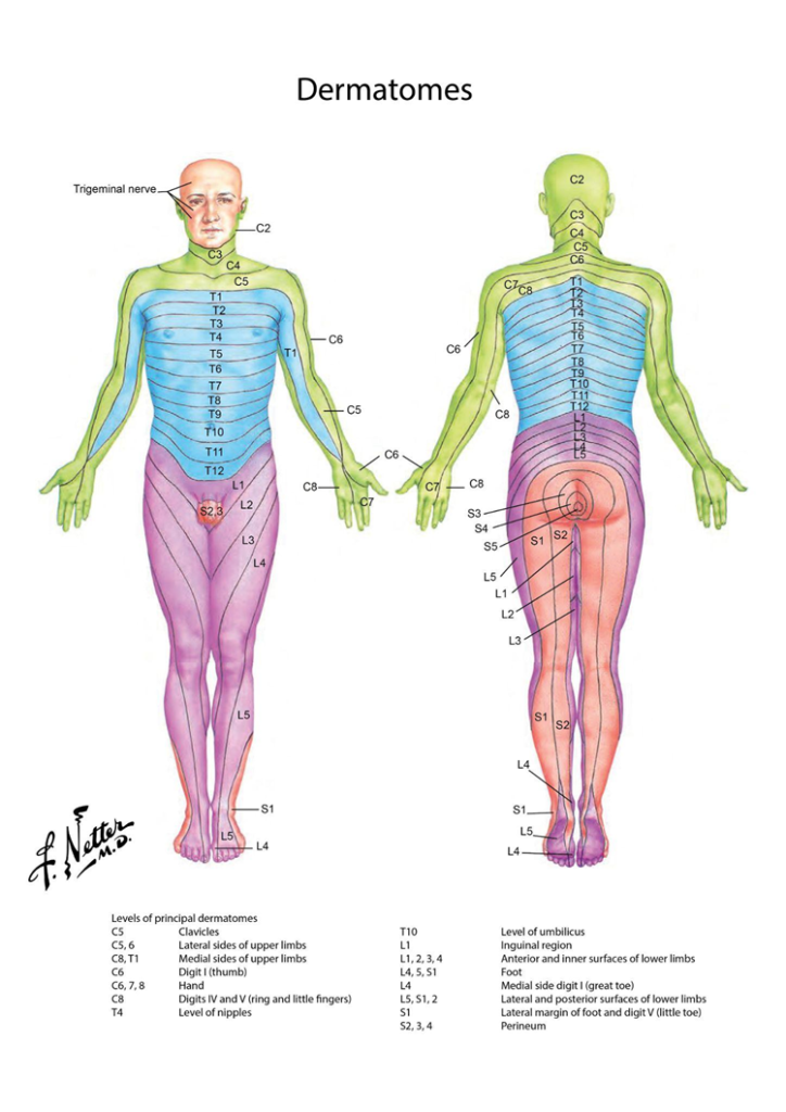
Dermatome Distribution Table
| Nerve Roots | Dermatom | ||
| Cervical | C2 | Innervation of the neck | Temples, Forehead, Back of Head |
| C3 | Entire Neck, Posterior Cheek, Temporal Area, Forward extension below Mandible | ||
| C4 | Shoulder Area, Clavicle Area, Upper Scapula Area | ||
| C5 | Innervation of the upper arm | Deltoid Area, Anterior aspect of the entire arm to the base of the thumb | |
| C6 | Anterior Arm, Radial side of the hand to the thumb and index finger | ||
| C7 | Lateral Arm and Forearm to the index finger, long finger, and ring finger | ||
| C8 | Medial Arm and forearm to long, ring, and little fingers | ||
| Thoracal | T1 | Medial side of the forearm to the base of the little finger | |
| T2 | Innervation of the chest and abdomen | Medial side of upper arm to medial elbow, chest and midscapular area | |
| T3 – T6 | Upper Thorax | ||
| T5 – T7 | Costae Margin | ||
| T8 – T12 | Abdomen and Lumbar Area | ||
| Lumbal | L1 | Return, past the trochanter and groin | |
| L2 | Back, front of thighs to knees | ||
| L3 | Innervation of the feet | Back, upper back, anterior thighs and knees, medial lower leg | |
| L4 | Medial buttocks, lateral thighs, medial legs, instep, big toe | ||
| L5 | Buttocks, posterior and lateral thighs, lateral aspect of the lower leg, instep, medial half of the foot, first, second, and third toes | ||
| Sacral | S1 | Buttocks, Thighs, and Posterior Legs | |
| S2 | Innervation of the groyne area | Buttocks, Thighs, and Posterior Legs | |
| S3 | Groin, medial thigh to knee | ||
| S4 | Perineum, genitals, lower sacrum | ||
Table of Major Dermatome Grades
| Nerve Roots | Tingkat Dermatom |
| C5 | Clavicle |
| C5 – C6 | Lateral side of the upper leg |
| C8 – T1 | Medial side of the upper leg |
| C6 | Thumb |
| C6 – C8 | Hand |
| C8 | 4th and 5th fingers |
| T4 | Nipple level |
| T10 | Navel level |
| L1 | Area inguinal |
| L1 – L4 | Front and inner side of the lower leg |
| L4 – S1 | Foot |
| L4 | Medial side of the big toe |
| L5 – S2 | lateral and posterior sides of the lower leg |
| S1 | Lateral border of the foot and toes 5 |
| S2 – S4 | Perineum |
Also read: Parkinson's: What is it and what is the role of physiotherapy
Reference :
- Whitman PA, Adigun OO. Anatomy, Skin, Dermatomes. [Updated 2022 Sep 12]. In: StatPearls [Internet]. Treasure Island (FL): StatPearls Publishing; 2023 Jan-. Available from: https://www.ncbi.nlm.nih.gov/books/NBK535401/
- Physiopedia. Dermatome. diakses pada tanggal 26 Oktober 2023 melalui https://www.physio-pedia.com/Dermatomes
- Netter, F. H. 1. (2014). Atlas of human anatomy. Sixth edition. Philadelphia, PA, Saunders/Elsevier.